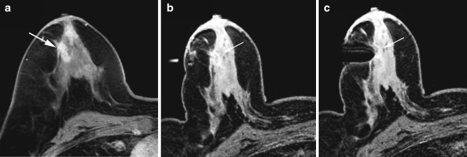
Fig. 3a–c.
Typical example of a biopsy procedure. a Diagnostic post-contrast T1-weighted image shows suspicious lesion. b Pre-biopsy image, compressed breast with guiding marker tube at the same location as the lesion. c The needle is inserted in front of the lesion; 3-5 samples were obtained. Histology: DCIS

