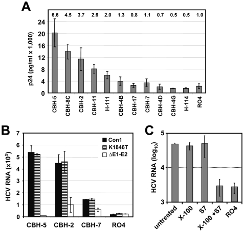
Figure 4. Capture of Con1 particles by E2-specific antibodies and characterization of captured particles.
(A) Identification of monoclonal antibodies reacting with Con1 envelope glycoproteins. HCVpp carrying Con1 envelope glycoproteins were incubated with immobilized antibodies specified in the bottom and captured particles were quantitated by using p24-specific ELISA. Numbers above each bar refer to fold increase above background as determined by capture assay with an irrelavant antibody (RO4). Mean values of three experiments and the standard deviations are given. (B) Capture of HCV particles from supernatants of Huh-7 cells transfected with Con1/wt or Con1/K1846T or the Con1 mutant lacking most of the envelope glycoprotein coding region (ΔE1–E2). Bound particles were quantified by using TaqMan qRT-PCR. Mean values of triplicate measurements including the standard error of the means are given. (C) Characterization of captured Con1/wt particles by nuclease treatment. Concentrated culture supernatant of Con1/wt transfected cells was used for capture with control antibody R04 (right bar) or CBH-5 (all other bars). Immune complexes obtained with the latter were split into 4 aliquots that were left untreated or treated with 0.5% Triton X-100, or nuclease S7, or 0.5% Triton X-100 and S7 nuclease. RNA was extracted and quantified by TaqMan qRT-PCR.

