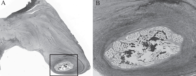
Figure 5).
A Histological section showing the synthetic chorda surrounded by tissue fibrosis (original magnification ×2.5; hematoxylin and eosin stain). B Higher magnification (box from A) showing intact synthetic chorda with collagen deposition in the interstices of the Dacron sutures. There was no evidence of chorda degradation. No inflammatory cells were seen (original magnification ×10; Movat pentachrome stain)

