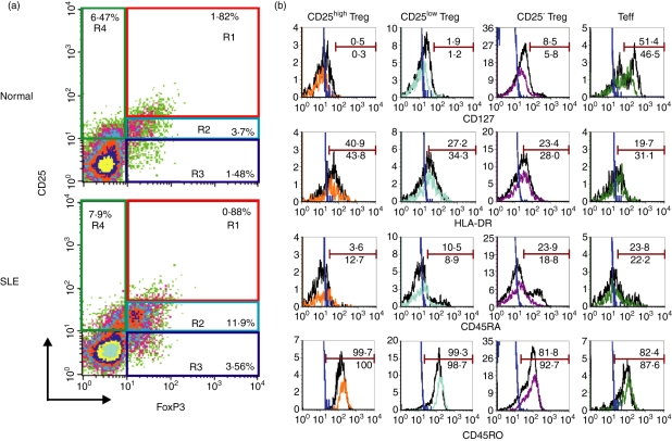
Figure 1.
Characterization of different subsets of regulatory T cells (Tregs) and effector T cells (Teffs) in patients with systemic lupus erythematosus (SLE) and in controls. (a) Purified T cells from peripheral blood mononuclear cells (PBMCs) were stained with monoclonal antibodies specific for CD4, CD25 and intracellular forkhead box P3 (FoxP3) and analyzed using a FACScan. Representative dot-plots show the expression of CD25 and FoxP3 on gated CD4+ T cells from one healthy donor (upper panel) and from one patient with SLE (lower panel). Quadrants were established using appropriate isotype controls. CD4+ CD25high FoxP3+ (R1), CD4+ CD25low FoxP3+ (R2) and CD4+ CD25− FoxP3+ (R3) cells were termed as CD25high Tregs, CD25low Tregs and CD25− Tregs, respectively. CD4+ CD25+ FoxP3− (R4) cells were termed as Teffs. (b) PBMCs were stained with monoclonal antibodies specific for CD4, CD25, CD127, HLA-DR, CD45RO, CD45RA and intracellular FoxP3 and analyzed using LSRII. Histograms show surface marker expression on different Treg subsets and Teffs of one healthy donor (black line) and one patient with SLE (color line). The blue lines represent the isotype control. Data are representative of five independent controls and four independent patients.

