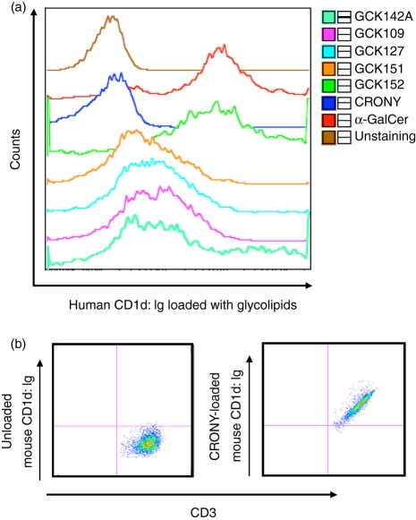
Figure 4.
Staining of invariant natural killer T (iNKT) cells with CD1d:Ig dimeric protein loaded with glycolipids was determined using FACS. (a) Approximately 2 × 10⁵ human iNKT cells were incubated for 1 hr at room temperature with 1 μg of a recombinant human CD1d:mouse Ig dimeric protein preloaded overnight with 0·5 μg of each glycolipid. The iNKT cells were then stained with phycoerythrin (PE)-conjugated anti-mouse IgG and analyzed by FACS. The results represent one of three similar experiments in which different human iNKT cell lines were generated from three different donors. (b) Approximately 2 × 10⁵ cells of murine iNKT hybridoma 1.2 cells were incubated for 1 hr at room temperature with 1 μg of a recombinant mouse CD1d:mouse Ig dimeric protein preloaded overnight with 0·5 μg of α-C-galactosylceramide (α-C-GalCer; CRONY). The hybridoma cells were then stained with allophycocyanin-conjugated anti-CD3 immunoglobulin IgG and with PE-conjugated anti-mouse IgG and analyzed by FACS. The results represent one of two similar experiments.

