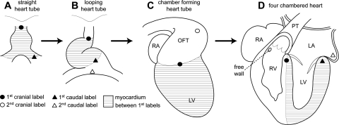
Fig. 3.
The fate of the early heart tube. A summary of experiments by De la Cruz et al. [12–14]. The early heart tube was labeled and reincubated. Ventral views of a straight (a) and a looping (b) heart tube. c Right view of a chamber-forming heart. d Ventral view of a four-chambered heart, showing the inflow of the left ventricle (LV) and the outflow of the right ventricle. LA left atrium, PT pulmonary trunk, RA right atrium, RV right ventricle

