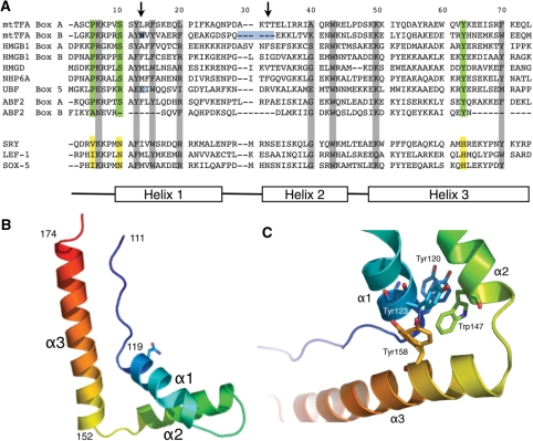
Figure 4.
X-ray crystal structure of human mtTFA box B. (A) Sequence alignments of box A and box B of h-mtTFA, box A and box B of HMG1, HMGD, NHP6A and Abf2. Global structure of an HMG protein is shown by diagram of helices α1, α3 and α3 at the bottom. Green boxes are residues that are conserved among all of the proteins. The arrows point to residues that are known to be involved in intercalating the DNA. (B) Ribbon diagram drawing of the backbone of box B showing the global fold of three helices stabilized into an L-shape configuration. The image was generated with PYMOL (40). (C) Hydrophobic core of box B with amino acids Tyr120, Tyr123, Trp147 and Tyr158 shown.

