Figure 8.

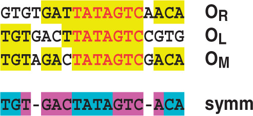
Comparison of operator sequences in Esp1396I. C.Esp1396I binding sites at the CR promoter (proximal, OR; distal, OL) and the M promoter (OM) are aligned. Indicated in yellow are the sites where at least 2/3 of the sequences are identical, seven of which (red lettering) are found in all three. A symmetrical consensus sequence is shown below, closely matching the OM site. Bases identified from the crystal structure as being involved in DNA–protein interactions are indicated in blue; other symmetrical bases likely to interact with helix 3 of the protein are shown in magenta.
