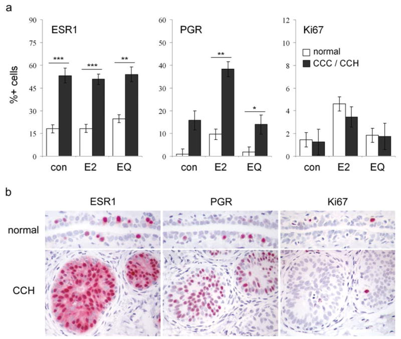
Figure 4.

Immunolabeling of columnar cell lesions for estrogen receptor alpha (ESR1), progesterone receptor (PGR), and the proliferation marker Ki67. (a), labeling indices across treatments, stratified by lesion type. CCLs included columnar cell change (CCC) and columnar cell hyperplasia (CCH) lesions. Vertical bars indicate standard errors. * P < 0.05, ** P < 0.01, and *** P < 0.001 compared to respective normal epithelium values by Wilcoxon Rank Sum test. (b), representative immunostaining of normal duct (top row) and CCH (bottom row) for ESR1, PGR, and Ki67 in an E2-treated animal.
