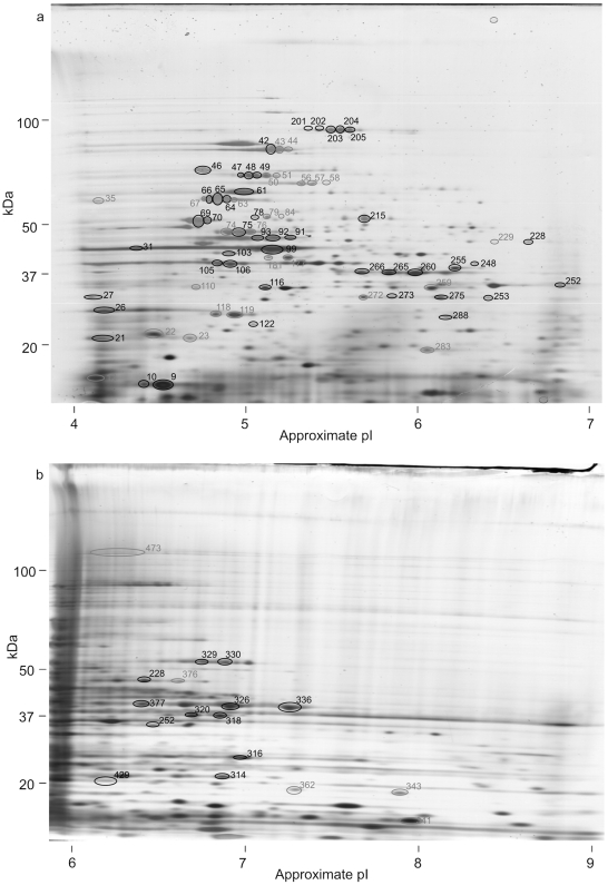
Figure 1. 2D gels of total N. meningitidis proteins.
Total N. meningitidis proteins separated by 2D gel electrophoresis using (a) a non-linear pI 4–7 1st dimension and (b) a non-linear pI 6–9 1st dimension. Gels were silver stained and replica gels western blotted with patient sera. Spots that were recognised by one or more sera on western blots are circled. Spots whose identity was determined are numbered in black, those that remain unidentified in grey.

