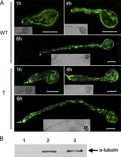
Fig. 7.
Subcellular localization of the α-tubulin protein in Arabidopsis pollen tubes. In vitro cultured pollen tubes were fixed, incubated with anti-α-tubulin antiserum, and detected with goat anti-mouse secondary antibody as described in the Materials and methods. The fluorescence of stained pollen tubes was observed under a confocal microscope. (A) Fluorescence from immunolabelling of Arabidopsis pollen tubes harvested at 1, 4, and 6 h after germination. WT indicates pollen of wild-type Arabidopsis; T indicates pollen of transgenic Arabidopsis. The transgenic tubes tip (arrows) were strongly labelled with antibody for α-tubulin, whereas wild-type pollen tubes showed a much weaker signal in the apical region (asterisks). These data were obtained from three independent experiments, and every condition was tested three times. Bars=20 μm. (B) Western blot of extracts (equal loading of samples) from Arabidopsis pollen tubes (lane 2) and P. wilsonii pollen tubes (lane 3) probed with anti-α-tubulin antibody. Lane 1 is the blank control.

