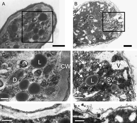
Fig. 8.
Effects of PwTUA1 on the ultrastructure of pollen tubes in transgenic Arabidopsis. Pollen from wild-type and transgenic plants was cultured in vitro for 6 h. Bars=0.2 μm (A, B, E, F) or 0.5 μm (C, D). (A) Tip region of wild-type pollen tube showing organelles at the tip. (B) Tip region of a pollen tube expressing PwTUA1; MTs (arrowhead) appeared and the number of vacuoles and secretory vesicles increased. (C and D) Magnified images of boxed sections in A and B. (E) Pollen tube wall of wild-type Arabidopsis. (F) Pollen tube wall of transgenic Arabidopsis. For Arabidopsis expressing PwTUA1, analyses of two independent transgenic lines showed a similar ultrastructure of pollen tubes. Data from one transgenic line were used. CW, cell wall; Mi, mitochondria; D, dictyosome; SV, secretory vesicle; V, vacuole; L, lipid body.

