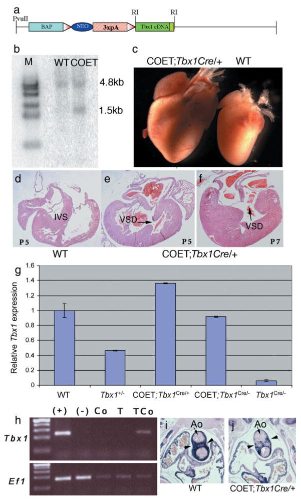
FIG. 1.
Generation of the COET transgene and COET transgenic mice. (a) Schematic of the COET construct; BAP: β-actin promoter, 3xpA: three repetitions of a polyadenylation signal; triangles, loxP; RI: EcoRI. (b) Southern blot autoradiography confirms integration of the entire transgene into the ES cell clone used for the generation of the transgenic line. A Tbx1 exon 1 specific probe reveals a 4.8kb fragment from the endogenous allele and a 1.5kb transgenic fragment. Genomic DNA was cut with EcoRI. (c) Hearts dissected from 7-day-old littermates. The hearts of COET;Tbx1Cre/+ are greatly enlarged. (d–f) Transverse sections of P5 and P7 animals. Note the thickened myocardium and VSD in the COET;Tbx1Cre/+ hearts. IVS, interventricular septum. (g) Quantification of Tbx1 expression. Quantitative real-time PCR analysis of the cranial region of 3 mutants per genotype shows that Tbx1 dosage from the COET transgene corresponds to about 1.8x Tbx1. At E10.5 one activated transgene expresses approximately much Tbx1 as 2 wild-type alleles. (h) Reverse transcription PCR of RNA samples from a whole E10.5 wild type embryo (+), Tbx1−/− embryo (−), dissected heart from a E14.5 COET embryo (Co), a Tbx1Cre/+ embryo (T), and a COET;Tbx1Cre/+ embryo (TCo). Note that Tbx1 expression is only detectable in the heart of the latter embryo. Ef1 is a positive control carried out in the same samples. (i,j) Coronal sections from E12.5 embryos (at the level of the cardiac outflow tract) immunostained with an anti α smooth muscle actin antibody. Note the much lighter staining of the outflow tract (arrowheads) in the conditional expression mutant (j) as compared with the wild type control (i). Ao: aortic valve.

