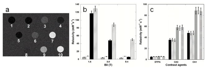
Figure 2.
Comparison of in vitro relaxivity between DTPA and designed contrast agents. (a) MR images produced using an inversion recovery sequence (TR 6000 ms, TI 960 ms, and TE 7.6 ms) at 3T. Samples are 1) dH2O, 2) 10 mM Tris-HCl pH 7.4, 3) 0.10 mM Gd3+-DTPA in H2O, 4) 0.10 mM Gd3+-DTPA in 10 mM Tris-HCl pH 7.4, 5) 0.10 mM Gd3+ and CD2, 6) 0.077 mM Gd3+-CA4.CD2, 7) 0.050 mM Gd3+-CA2.CD2, 8) 0.10 mM Gd3+-CA9.CD2, 9) 0.020 mM Gd3+-CA1.CD2, and 10) 0.050 mM Gd3+-CA1.CD2. (b) Proton relaxivity values of Gd3+-CA1.CD2 (r1, solid black; r2, cross) and Gd3+-DPTA (r1, shield; r2, open) at indicated field strength were measured as a function of field strength. (c) In vitro relaxivity of contrast agents Gd3+-DPTA (DTPA), Gd3+-CA1.CD2 (CA1) and Gd3+-CA2.CD2 (CA2) in the absence of Ca2+ (black and grey), presence of 1 mM Ca2+ (left strip and open) and 10 mM Ca2+ (right strip and cross) at 3T. T1 (black, left & right strips) and T2 (grey, open and cross) were determined using a Siemens whole-body MR system.

