
Fig. 9. ADAR2 protein expression in mouse brain at P0.
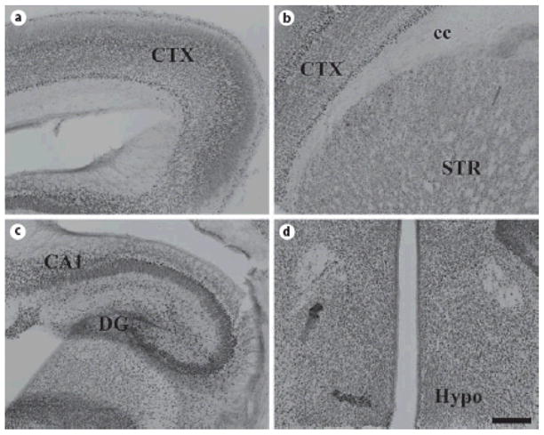
Coronal sections of P0 mouse brain were stained for ADAR2. Similar to ADAR1, widespread expression in deep and superficial layers of the still developing cerebral cortex (A, B), hippocampus (C), thalamus (D), and hypothalamus (D) is readily observed. Labeling in the striatum is fairly low (B). Scale bar = 200 μm.