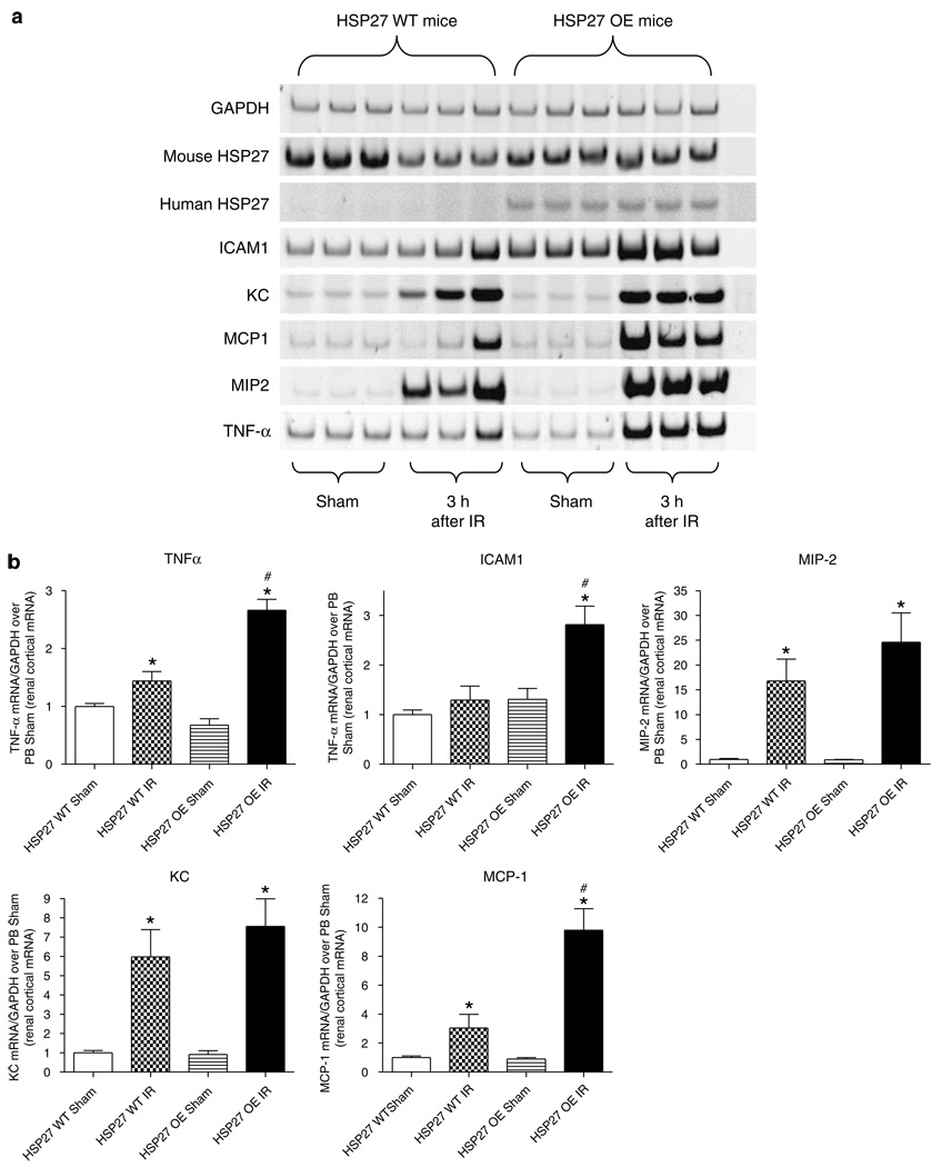
Figure 5. HSP27 OE mice show reduced proinflammatory mRNA expression after renal IR.
(a) Representative gel images of semiquantitative RT-PCR of mouse and human HSP27 as well as proinflammatory markers ICAM-1, KC, MCP-1, MIP-2, and TNF-α from renal cortices of HSP27 WT and huHSP27 OE mice subjected to sham operation or to renal ischemia and 3 h reperfusion. (b) Densitometric quantifications of relative band intensities normalized to GAPDH from RT-PCR reactions for each indicated mRNA. *P < 0.05 vs appropriate sham. #P < 0.05 vs HSP27 WT IR. Error bars represent 1 s.e.m.

