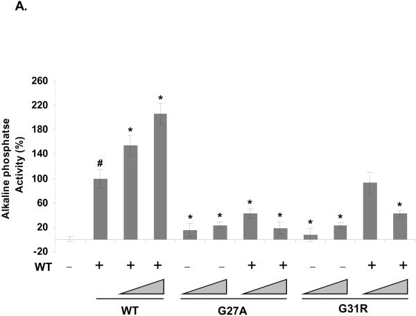
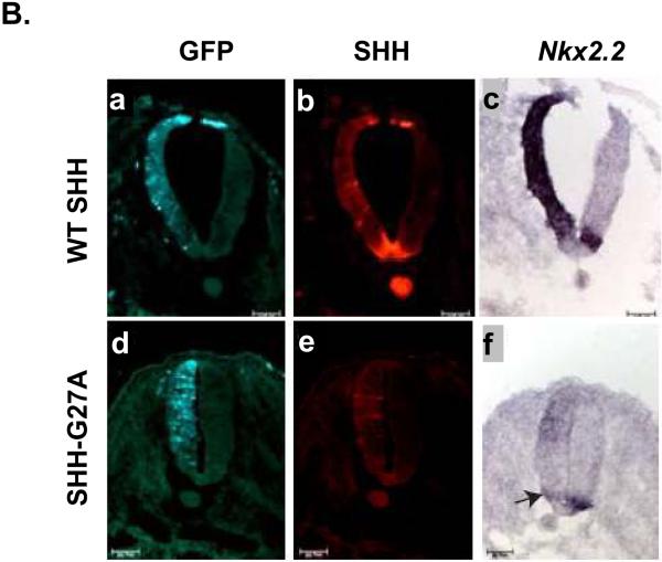
Figure 3. SHH-G27A dominantly inhibits endogenous SHH activity.
The ability of SHH mutants SHH-G27A and SHH-G31R to affect wt SHH activity was evaluated by testing conditioned medium from Bosc cells expressing wt SHH (WT), SHH-G27A (G27A) and SHH-G31R (G31R) alone or in combination with wt SHH, at different molar ratios (1x:1x and 5x:1x respectively), for its ability to induce alkaline phosphatase activity in C3H10T½ cells (A). In this experiment, 1x corresponded to 80 ng of variant DNA and a total of 500 ng of DNA (equalized using vector DNA) was transfected into Bosc cells plated in 60 mm plates. Activity assays were done in triplicate and the error bars represent ±S.D. Both SHHG27A and SHH-G31R attenuated wt SHH activity (1x wt SHH, indicated by #) in a dose dependent manner. The activity of these mutants alone or when expressed in combination with wt SHH was significantly different from 1x wt SHH as were higher doses of wt SHH (P values < 0.05 are indicated by *). SHH-G27A also attenuated endogenous SHH activity in vivo (B). The ability of SHH-G27A to attenuate endogenous SHH in the chick neural tube was assessed by electroporating a SHH-G27A expressing plasmid (n=16), along with a GFP expressing plasmid, on one side of the chick embryonic neural tube. SHH expression was determined using anti-SHH antibodies (red). Induction of Nkx2.2 expression (dark blue) was determined by in situ hybridization. GFP expression (light-green) served as an electroporation control in these experiments. SHH-G27A attenuated the induction of Nkx2.2 expression by endogenous SHH (arrow in panel f) validating our in vitro observation that some SHH mutants can act in a dominant negative manner. Under these conditions, endogenous SHH activity was attenuated 50% (8 out of 16) of the time embryos were electroporated with SHH-G27A expressing plasmid (see Table 1).


