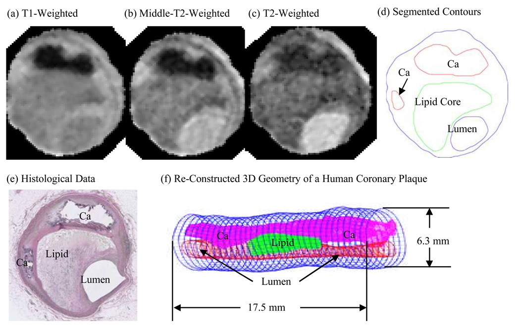
Figure 1.
A human coronary atherosclerotic plaque sample: Multi-contract MR images and re-constructed 3D geometry. (a)–(c), MR images with T1, middle-T2, and T2-weighted images. (d) Contour plot of the segmented image using a multi-contrast algorithm; (e) Histological data. The location and shape of each major plaque component correlated very well with histological data; (f) 3D plaque geometry re-constructed from a 36-slice ex vivo MRI data set.

