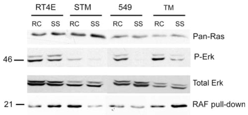
Fig. 6.

Ras is constitutively activated in 2 MRT cell lines. 30 μg of cell lysates from randomly cycling (RC) and serum-starved (SS) RT4E, STM91-01 (STM), TTC549 (549), and TM87-16 (TM) cell lines were separated by 10% SDS-PAGE. Western blotting was performed by cutting the membrane in half and probing the bottom half with a pan-Ras antibody (top panel) and the top half with an antibody to phosphorylated Erk (P-Erk) (second panel) followed by antibody to total Erk (third panel). Cell lysates of 200 μg were also immunoprecipitated using Raf-1 RBD agarose beads. SDS-PAGE of 10% was used to separate immunoprecipitation reactions and Western blotting was performed with a pan-Ras antibody (bottom panel)
