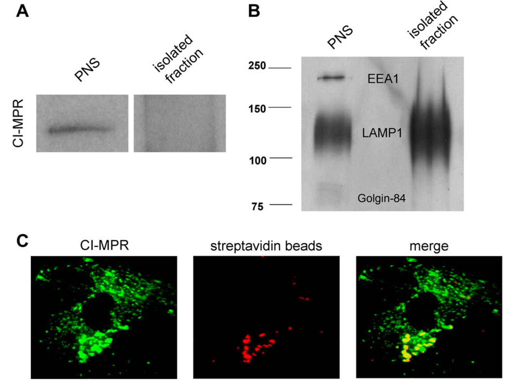
Fig. 2.
Western blot and immunofluorescence analysis shows intracellular localization of the FRET probes in two biochemically distinct compartments. Using optimized pulse–chase conditions, FRET probes were endocytosed into cells and their specific organelle localization was analyzed. (A) Magnetic isolation of lysosomes using iron-coated dextran of the same size was performed as described previously [7]. Western blot analysis was performed using antibodies specific to the CI-MPR. (B) Golgin-84, EEA1, and LAMP1 on fractions of PNS and isolated lysosomes. The isolated lysosome fraction was found to be devoid of late endosome, early endosome, and Golgi protein markers and enriched in LAMP1. (C) Immunofluorescence was performed after endocytosis of streptavidin beads (red). The beads were found to localize in CI-MPR-positive (green) compartments. (For interpretation of the references to color in this figure legend, the reader is referred to the Web version of this article.)

