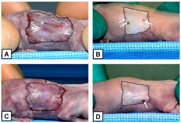
Figure 5.
Representative images, at 2 and 6 weeks after surgery, of athymic mice grafted with ESS fabricated with keratinocytes harvested from Kerator (A and B) and flasks (C and D). The perimeter of grafted area has been delineated with dashed lines. Note the extent of wound contraction in both conditions. Also remarkable is the presence of pigmented spots within the grafted area at 6 weeks in both conditions (arrows), suggesting the presence of human derived cells in those regions.

