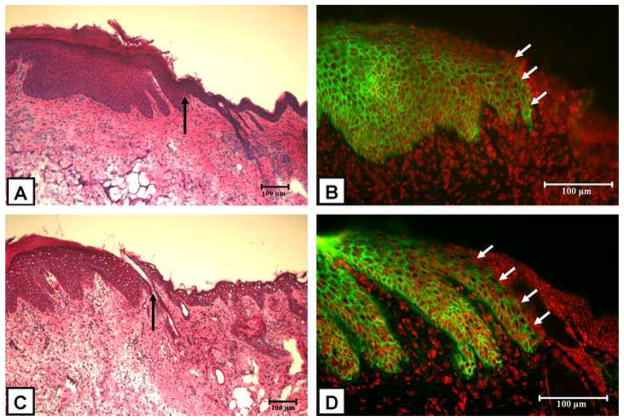
Figure 7.
Representative H&E stained sections and HLA-ABC immunofluorescence of grafted ESS fabricated with keratinocytes harvested from Kerator (A and B, respectively) and flasks (C and D, respectively). In both conditions, H&E sections (A and C) show normal mouse epidermis (to the right of arrow) which is thin and lacks rete ridges. The thicker epithelium with prominent rete ridges (to the left of arrow) resembles human skin and is derived from the grafted ESS. Direct immunofluoresence for HLA-ABC antigens (B and D) shows net-like distribution of HLA-ABC (green) on human derived keratinocytes in the epidermis. Adjacent mouse epidermis (arrow) stains negatively for HLA-ABC. Nuclei have been stained red by propidium iodide. Scale bar = 100μm.

