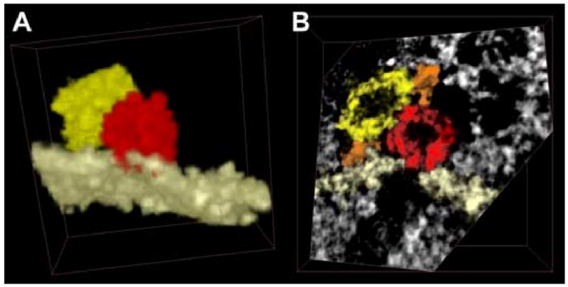
Figure 6.

A vesicle is fused with the plasma membrane and in contact with another vesicle. (a) After segmentation, everything was excluded from the image apart from a single membrane, the fused vesicle and the neighboring one. Though vesicles have often been represented with the same color (yellow), the segmentation process can assign a different label to each vesicle, allowing us to represent them with multiple colors when necessary (as done here). Note the red spot on the membrane, demonstrative of the fusion between vesicle and membrane. In (b) a single plane is shown, cut obliquely to the edge of the volume, for a better representation of the region where the two vesicles touch other. The plasma membrane is shown in white and two filaments in orange.
