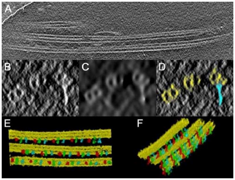
Figure 7.

Segmentation of a cryotomogram containing ciliary MT-doublets, obtained in single axis geometry. (a) A plane section along XY; the tilt axis is oriented parallel to the doublets. (b-d) a section orthogonal to the tilt axis direction: (b) original plane; (c) after non linear anisotropic diffusion; (d) after segmentation. MT-doublets are in yellow, and radial spokes in light blue. (e-f) volume rendering views of the segmented doublets with radial spokes attached. Three different colors (red, green and blue) have been assigned to each non equivalent radial spoke of a triplet.
