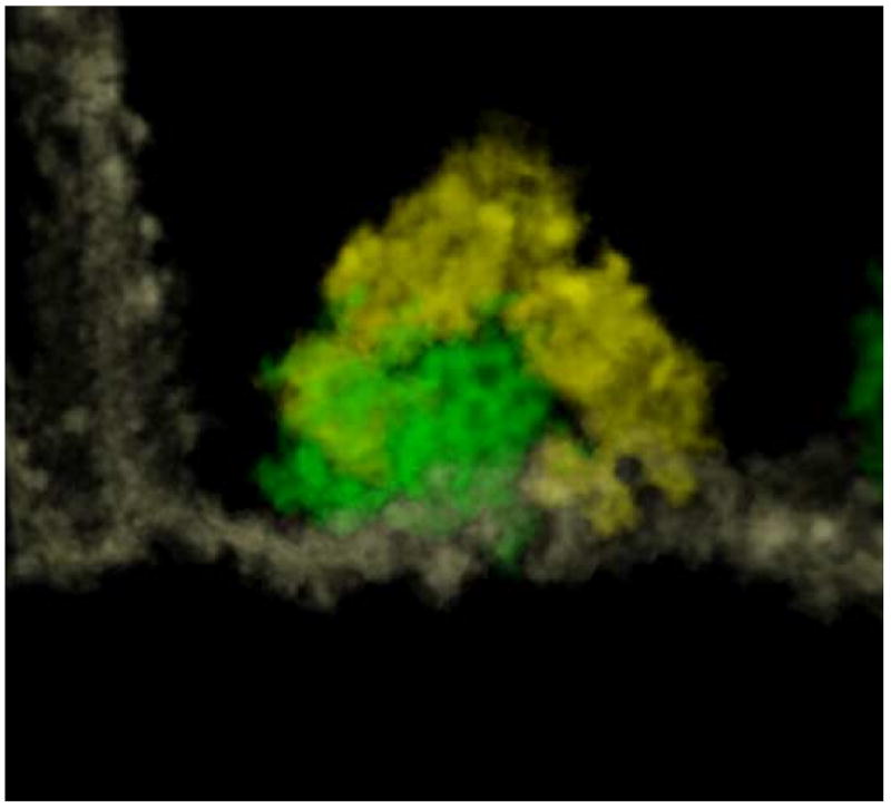
Figure 8.

A syndesome (green) has been segmented and visualized with a volume rendering technique, together with the portion of membrane (white) to which it is attached. The density of the structure is such that a cage with polygonal faces is quite well-defined. Vesicles surrounding the syndesome are evidenced in yellow.
