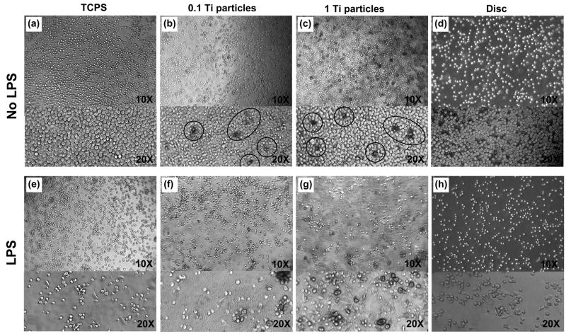
Figure 3.
Phage contrast cell images of THP-1 cells cultured for 72 hrs on TCPS (a and e), Ti particles at 0.1 mg/mL (b and f) and at 1 mg/mL (c and g), and Ti discs (d and h). The top panel(a, b, c and d) shows unstimulated cells and the bottom panel (e, f, g and h) shows LPS-stimulated cells (1 μg/mL LPS). Circles in the (b) and (c) demonstrate the blackened THP-1 cells resulted from Ti partilce engulfment. Note that cell density of images may not be representative because the cell distribution of suspended THP-1 cells in the rounded culture wells are not homogeneous along the substrates mainly caused by centrifuge force.

