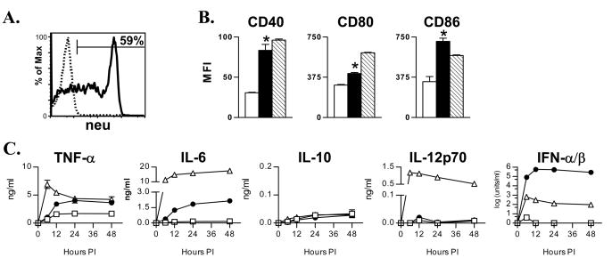
Figure 1. VRP transduction results in high-level transgene expression and DC activation.
A. DCs were transduced with neuET-VRP (MOI = 10) and analyzed 12 hours later for expression of neuET by FACS. B. DCs were mock-infected (open bars), infected with GFP-VRP (solid bars), or treated with 100 ng/ml LPS (hatched bars). 24 hours later, DCs were stained for CD40, CD80, and CD86. Expression of costimulatory molecules on VRP-infected DCs was determined by gating on GFP+ DCs. Bars represent the mean +/− SEM (n = 4 per group). One of four similar experiments is depicted. C. DCs were mock-infected (open squares), infected with GFP-VRP (closed circles), or treated with 100 ng/ml LPS (open triangles). Cytokine levels were evaluated at the indicated times post-infection as described in Materials and Methods. *p<0.001 v. mock-DCs, Student’s t test.

