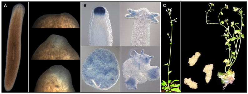
Figure 4. Invertebrate and Plant Model Systems for Studying Regeneration.
(A) The planarian Schmidtea mediterranea (left) and a regeneration series of the anterior end at ~days 1, 3, and 4 after decapitation.
(B) Head regeneration in Hydra after decapitation (top) and from a cell aggregate of cells obtained after dissociation of a Hydra into individual cells (bottom). The signal corresponds to the expression of a Chordin-like gene in this organism. Images reproduced from Rentzsch et al. (2007), Proc. Natl. Acad. Sci. USA 104, 3249–3254. Copyright (2007) National Academy of Sciences, USA.
(C) Normally developing Arabidopsis with a basal rosette and a reproductive inflorescence that has arisen from the transition of the SAM to a floral meristem (left). Middle, dedifferentiated cell masses of callus that formed from auxin treatment of tissue cuttings from Arabidopsis and the regeneration of a complete shoot from one such callus (right). Images of the callus and regenerating shoot are courtesy of S.P. Gordon and E.M. Meyerowitz.

