
Figure 2.
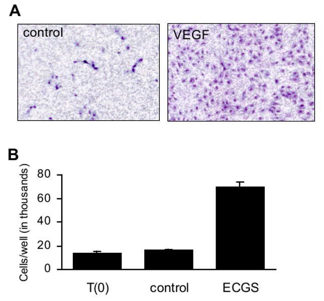
Cell culture assays assessing endothelial cell function. A) Cell migration as measured using a transwell assay. Starved HUVEC were plated onto a fibronectin-coated transwell with pores 3 μm pore diameter. Medium on the opposing side of the membrane lacked (left panel) or contained (right panel) recombinant VEGF at 10 ng/ml. After 18 hr, nonmigrated cells were removed with a cotton swab. Migrated cells were fixed and stained with hematoxylin and eosin, then photographed using a 5× objective. Unpublished images courtesy of Magali Saint-Geniez. B) Cell proliferation as measured by direct cell counting. HUVEC were treated with endothelial cell growth supplement and cultured for three days. The mean numbers of cells present in each well prior to the start of treatment (T(0)) and following three days of treatment (control, VEGF) are shown. Error bars indicate standard deviation. Unpublished data courtesy of Sandie Smith.