
Figure 4.
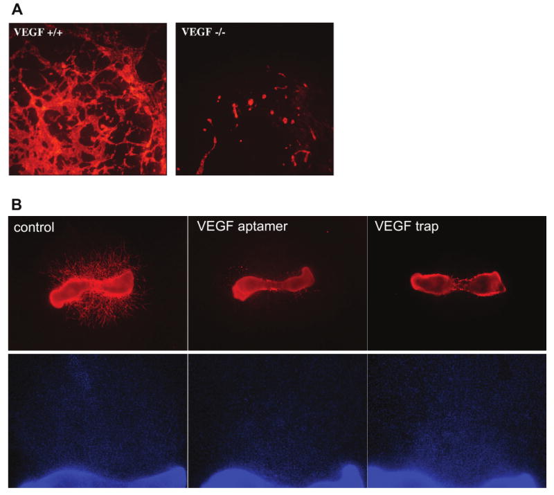
Embryoid body and mouse metatarsal models of angiogenesis. A) Vascular structures in embryoid bodies. Embryoid bodies were differentiated for 10 days from VEGF-deficient (left panel) or wild-type (right panel) mouse embryonic stem cells. The vascular structures were labeled using an antibody to PECAM and photographed at using a 5× objective. Unpublished images courtesy of Robyn Loureiro. B) Vessel outgrowth from mouse metatarsal bones. Metatarsal bones were isolated from wild type embryonic day 18 mouse embryos and cultured with control medium (left panels), an aptamer to VEGF (Ruckman et al., 1998; middle panels) or a soluble, truncated VEGF receptor (VEGF trap; Holash et al., 1992; right panels). Vessel outgrowth in the top panels is visualized by fluorescently labeling the endothelial cell marker PECAM and photographing using a 2× objective. The DAPI staining in the lower panels, photographed using a 4× objective, shows that the inhibitors do not prevent outgrowth of non-endothelial cells from the metatarsal bone. Unpublished images courtesy of Yin-Shan Ng.