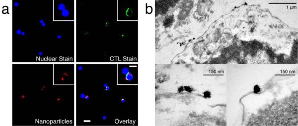
Figure 3.
Micrographs of targeting nanoparticle-labeled CTLs. (a) Fluorescently-labeled CTLs incubated with nanoparticles coupled with Alexa fluorophore (red). The cells were labeled with a DAPI for nuclear stain (blue) and with a FITC-CD8+ antibody for CTL identification (green). (b) TEM micrograph of CTLs labeled with targeting nanoparticles. Nanoparticles are shown, bound at the surface of the T cell cross sections.

