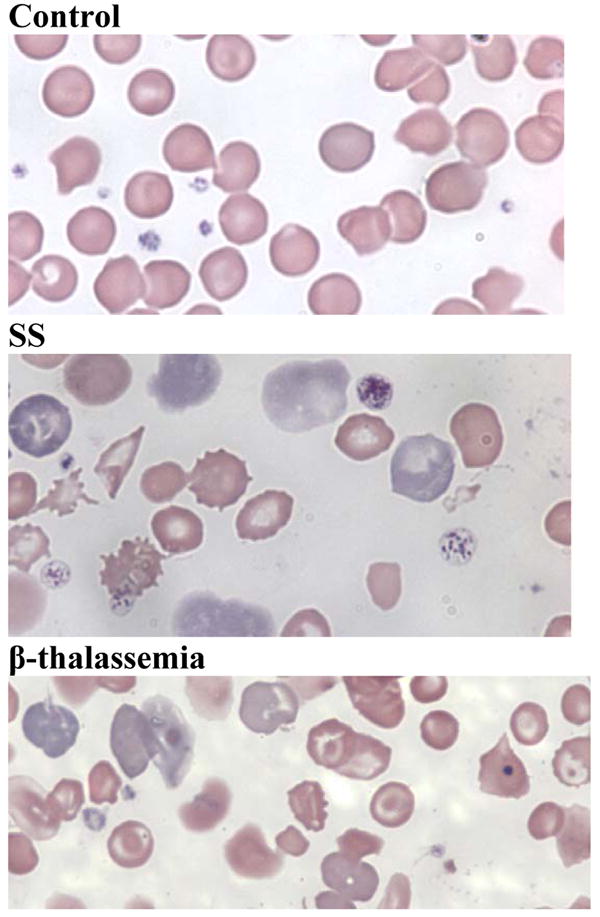
Figure 1. SS mice have markedly abnormal blood smears.

Peripheral blood smears of EDTA-anticoagulated blood obtained by retrobulbar venous plexus puncture from WT, SS and β-thalassemia mice. Blood smears from SS animals show anisocytosis, poikilocytosis, sickled erythrocytes, target cells, erythrocyte fragments of varying sizes, Howell-Jolly bodies, and an increased percentage of polychromatophilic erythrocytes. In sharp contrast, smears from WT animals do not show any of these findings. Blood smears from β-thalassemia mice show anisocytosis, microcytosis, targets cells, erythrocyte fragments, and an increased percentage of polychromatophilic erythrocytes.
