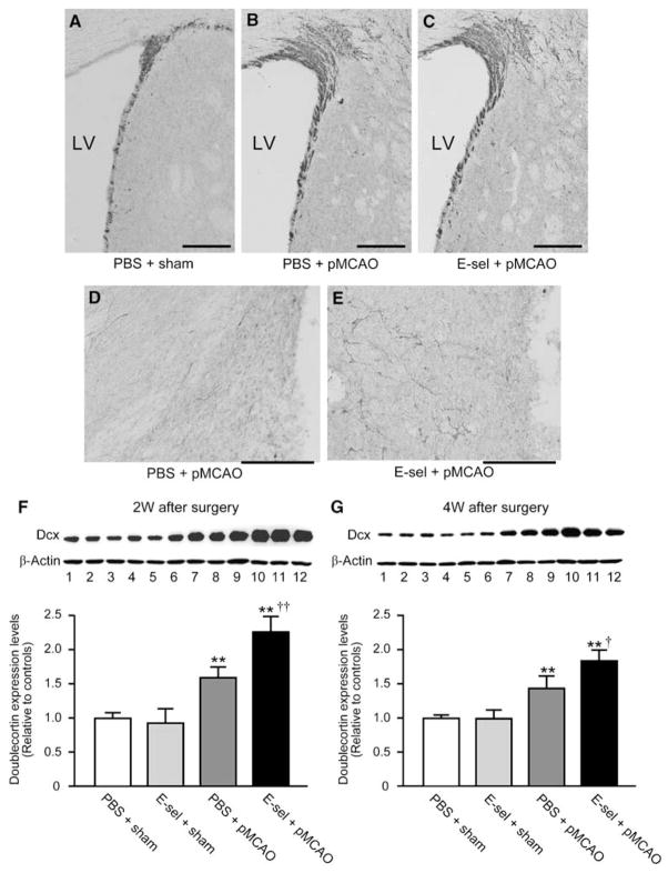
Figure 4.
Dcx protein levels were increased in the ischemic hemisphere of E-selectin-tolerized animals. Photomicrographs of the immunohistochemical staining for Dcx antibody in the ipsilateral SVZ (A–C) and periinfarct region (D, E) at 2 weeks after surgery. The cavity as shown on the right side of photomicrographs (D, E) is infarction. Scale bar=100 μm in A–E. LV=lateral ventricle. Representative western blot of doublecortin levels in brains exposed to PBS/sham, E-selectin/sham, PBS/pMCAO, or E-selectin/pMCAO (lanes 1 to 3, 4 to 6, 7 to 9, and 10 to 12, respectively) at 2 weeks (F) and 4 weeks (G) after surgery. Reblotting with anti-β-actin was performed as a loading control. Comparison of quantitative densitometric analyses of doublecortin protein levels in the brains. (n=4 for each group; *P<0.05, **P<0.01, compared with PBS+sham values, and †P<0.05, ††P<0.01, compared between PBS+pMCAO versus E-sel+pMCAO values; One way ANOVA followed by Bonferroni/Dunn post hoc test).

