
Fig. 3.
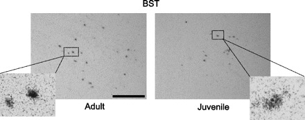
Photomicrographs showing representative regions of AVP mRNA-expressing cells in the BST in gonadally intact adult (left) and juvenile (right) rats. Cells were considered positive for AVP when the number of silver grains on top of the cells was 3 times higher than background. Insets show individual cells at higher magnification (×40). Scale bar, 10 μm.