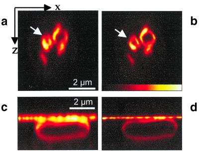
Figure 4.
Resolution improvement in live cells. XZ-images of a S. cerevisiae yeast cell with labeled vacuolar membranes with standard confocal resolution (a) and with axial resolution improved by STED (b). Whereas the confocal mode fails in resolving the membrane of small vacuoles, the STED microscopy better reveals their spherical structure. XZ-images of membrane-labeled E. coli show a 3-fold improvement of axial resolution by STED in d as compared with their simultaneously recorded confocal counterparts in c.

