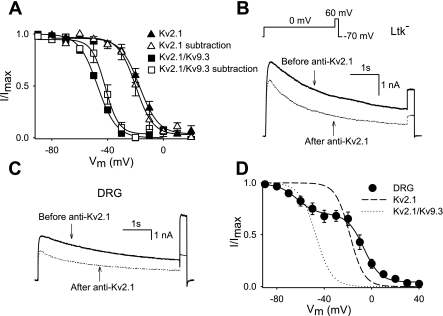
Fig. 4.
Voltage dependent activation and inactivation of the anti-Kv2.1-sensitive current. A: voltage-dependent inactivation of the anti-Kv2.1-sensitive current of Kv2.1 or Kv2.1/Kv9.3 heterologous expressed. The inactivation curve was obtained as in Fig. 3H. For comparison, the inactivation curves of Kv2.1 and Kv2.1/Kv9.3 obtained from heterologous expression are also shown. B: outward K+ current traces of Kv2.1/Kv9.3 expressed in Ltk− cells before (solid line) and after (dotted lines) diffusion of Kv2.1 antibodies into the cell. The voltage protocol is shown in the inset. C: representative current tracings of the outward K+ current of DRG neurons before (solid line) and after (dotted lines) intracellular diffusion of Kv2.1 antibodies. The same voltage protocol as in B was used. D: inactivation curve of the anti-Kv2.1-sensitive current was obtained as in Fig. 3H. Data were fitted with a double Boltzmann function (solid line). The inactivation curves of wild-type Kv2.1 and wild-type Kv2.1/Kv9.3 expressed in Ltk− cells are also shown. The midpoints of inactivation and slope factors are shown in Table 1.

