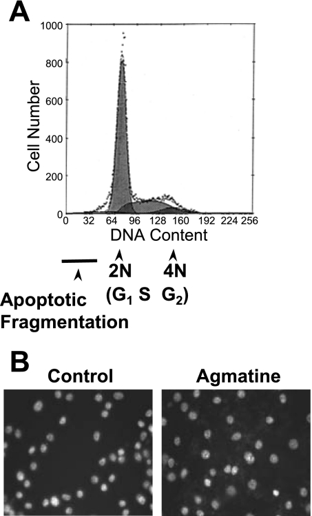
Fig. 1.
Apoptotic changes in DNA. A: propidium iodine/FACS analysis did not reveal an asymmetric shoulder to the left side of the 2N peak indicative of apoptotic DNA fragmentation. B: Hoechst staining of Ha-Ras-transformed NIH-3T3 (Ras/3T3) cells did not exhibit chromatin condensation typical of apoptosis. In both procedures, cells were incubated with 1 mM agmatine (AGM) for 10 days.

