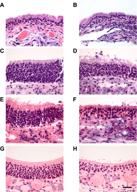
Fig. 2.
Histological sections of murine nasal airway. A: ciliated nasopharyngeal epithelia from WT mouse. B: CF nasopharyngeal ciliated epithelium. C: OE from 30-day-old WT mouse. D: OE from 30-day-old CF mouse. E: OE from 7-mo-old WT mouse. F: OE from 7-mo-old CF mouse. (Preparations in A–F were fixed after removal from Ussing chamber.) G: OE from 30-day-old CF mouse removed from nontreated side of mouse 5 days after undergoing olfactory bulbectomy (OBX) surgery. H: OE removed from the side receiving OBX surgery; same mouse as in G. All preparations were stained with hematoxylin and eosin. Size bar in H = 25 μM; magnification of all other sections is identical.

