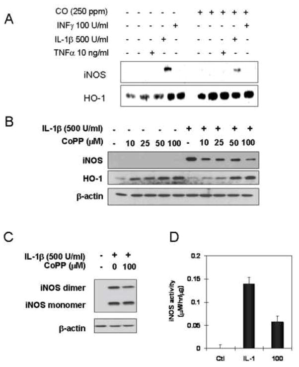
Figure 3.
HO-1 induction by CoPP suppresses the level of iNOS protein and active iNOS dimer in response to IL-1β. (A) Rat hepatocytes were cultured for 24h in the presence of cytokines (IL-1β;500 U/ml, INFγ;100U/ml, TNFα;10ng/ml) with or without CO (250 ppm) following the 2h pre-exposure of CO. Western blot for iNOS or HO-1 was performed using denaturing SDS-PAGE. (B). Cells were treated with CoPP at indicated concentration (10, 25, 50, 100 μM) for 20h and further incubated with treatment of IL-1β (500 U/ml) for 24h. Cytosolic fractions were prepared by centrifugation at 12,000×g for 20 min and western blot was performed using denaturing SDS-PAGE. (C). Cells were incubated with CoPP (100 βM) for 20h prior to addition of IL-1β (500 U/ml). After 12h cells were washed with ice-cold PBS, and lysed by three cycles of freeze and thaw. Cytosolic fractions were prepared by centrifugation at 12,000×g for 20 min and western blot was performed using non-denaturing SDS-PAGE. (D). Cytosolic iNOS activity was measured in the presence of 1 mM NADPH, 20 μM FAD, 20 μM FMN, and 4 mM L-arginine, with exogenous 100 μM BH4 at 37°C for 2h. Accumulated nitrite of cytosolic protein was measured using Griess reagent. Data represent the mean ± S.D. of more than three independent experiments

