Fig. 1.
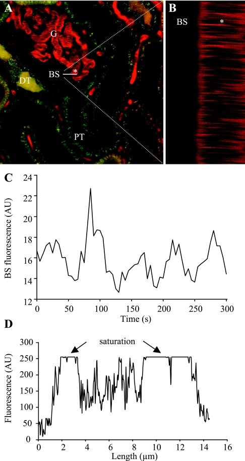
Two-photon imaging of glomerular permeability to macromolecules in the intact Munich-Wistar rat kidney in vivo. A: circulating plasma was labeled with 70-kDa dextran-rhodamine B (red), and proximal (PT) and distal (DT) renal tubules with quinacrine (green). G, glomerulus. B: line (xt) scan was performed at 1-ms intervals for 2 s along a horizontal line, ∼20 μm long as indicated on A. The left half of this line was positioned above the Bowman's space (BS) while the right half of it was over one glomerular capillary (indicated by *). Red blood cells appear as unstained dark objects (A) and leave dark bands on the line scan (B) as they move through the capillary (*). Intense plasma fluorescence appears between individual blood cells. C: recording of low levels of rhodamine B-fluorescence in the BS. Fluorescence intensity shows regular oscillations. D: linear profile of fluorescence intensities over the same glomerular capillary as selected in A, 2–14 μm indicate the outer diameter. At the edges, low-fluorescence signals are detected (capillary wall), while inside the capillary the plasma fluorescence is saturated (particularly in the relatively cell-free peripheral regions).

