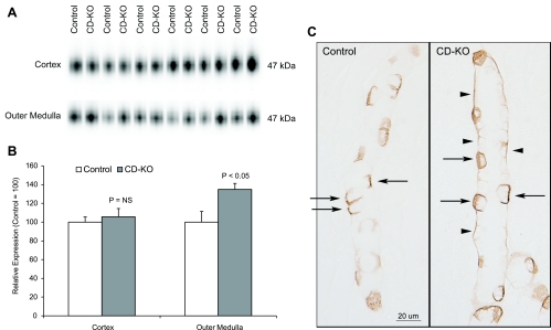
Fig. 7.
Rhbg expression in acid-loaded CD-KO and C mice. A: immunoblot analysis of Rhbg expression in the cortex and outer medulla. B: quantification of Rhbg protein expression. Cortical Rhbg expression is not significantly different between CD-KO and C mice undergoing HCl-induced metabolic acidosis. In the outer medulla, Rhbg expression is significantly greater in CD-KO mice than in C mice. C: Rhbg immunolabel in the OMCD of C and CD-KO mice. Basolateral Rhbg immunolabel in OMCD principal cells (arrowheads) is increased relative to immunolabel in acid-loaded C OMCD. Intercalated cell (arrows) basolateral Rhbg immunolabel appears unchanged.

