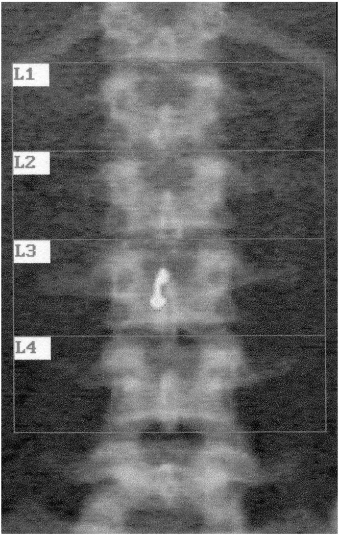
Figure 1.

Image from dual energy xray absorptiometry of lumbar spine in a subject wearing navel jewelry overlying the 3rd lumbar vertebra. The BMD was 0.909 g/cm2 initially. The jewel was traced and deleted from the analysis (“erased”). The area decreased by 0.66 cm2, and the new BMD was 0.887 g/cm2
