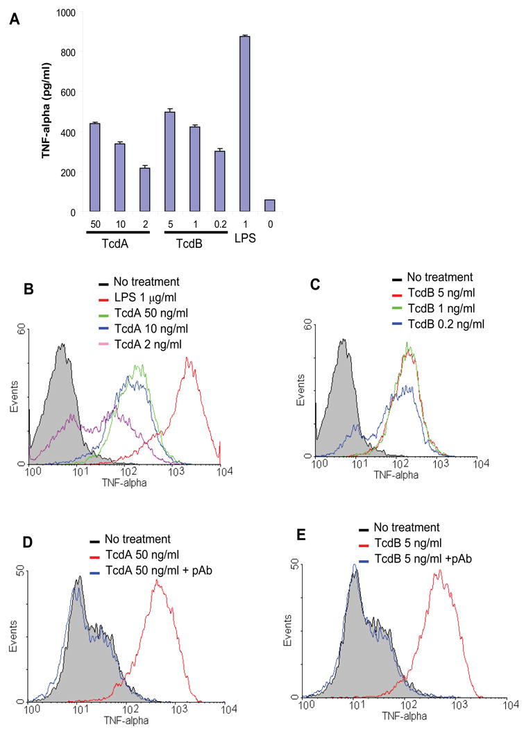
Fig. 1. TNF-α release by RAW 264.7 cells stimulated with TcdA or TcdB.

(A) RAW 264.7 cells were cultured for 18 h in the presence of TcdA or TcdB at the indicated doses (ng/ml). Control groups included LPS at 1 μg/ml and medium alone. TNF-α concentration in the medium was determined by ELISA. Data are mean ± SD (n=3). (B) RAW 264.7 cells were treated with TcdA at 50 ng/ml (green line), 10 ng/ml (blue line) or 2 ng/ml (purple line) for 6 h. (C) RAW 264.7 cells were treated with TcdB at 5 ng/ml (red line), 1 ng/ml (green line) or 0.2 ng/ml (blue line) for 6 h. Control groups include no treatment (grey line) or LPS (1 μg/ml, red line in (A)). (D and E) RAW 264.7 cells were treated with TcdA (50 ng/ml) (D) or TcdB (5 ng/ml) (E) for 6 h in the presence (blue line) or absence (red line) of a neutralizing antibody against both TcdA and TcdB. TNF-α was measured by an intracellular cytokine staining followed by flow cytometry analysis.
