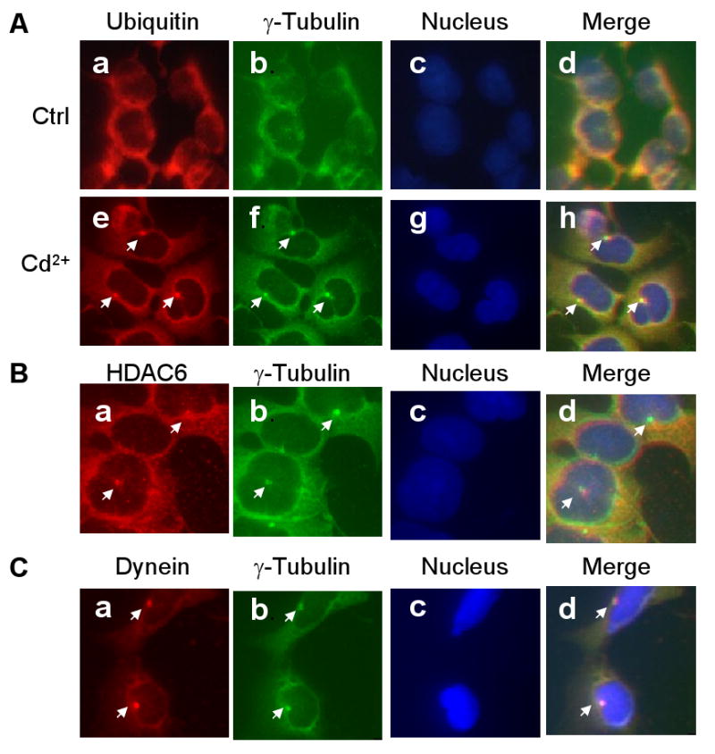
Fig. 1.

Cd2+-induced aggresome formation in HEK293 cells. HEK293 cells were cultured in the presence or absence of 20 μM Cd2+ for 6 h, then double-immunostained with antibodies against ubiquitin (Panel A, a, e; red) and γ-tubulin (Panel A, b, f; green). Panel B: Cd2+-treated cells immunostained with antibodies against HDAC6 (a, red) and γ-tubulin (b; green). Panel C: Cd2+-treated cells were immunostained with antibodies against dynein (a, red) and γ-tubulin (b, green). The nuclei (Panel A, c, g; Panel B, c; Panel C, c) were stained with DAPI (blue). Merged immunostaining images are shown in A, d, h; B, d; and C, d.
