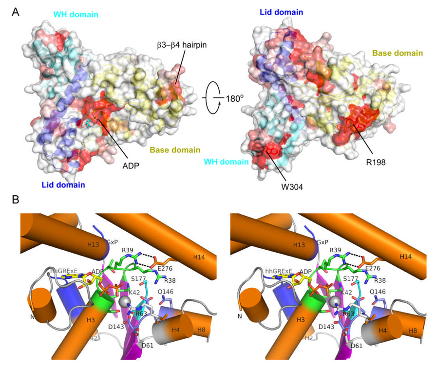
Figure 2.
Surface conservation pattern and the nucleotide binding site of SSO1545. (A). Molecular surface of SSO1545 colored by sequence conservation. The most conserved residues are shown in red, the non-conserved residues in white. The three domains of SSO1543 are shown in ribbon representation and colored as yellow, blue and cyan respectively. The orientation of left panel is the same as in Fig. 1A. (B) Close-up stereo view of the ADP binding site. The bound ADP (yellow) and magnesium ion (silver) are shown in sticks and sphere respectively. Walker A (P-loop, green), Walker B (W-B, blue), sensor I (S-I, cyan), sensor II (S-II, white) are shown in cartoon and sticks. The STAND specific hhGRExE and GxP motifs are also highlighted in blue.

