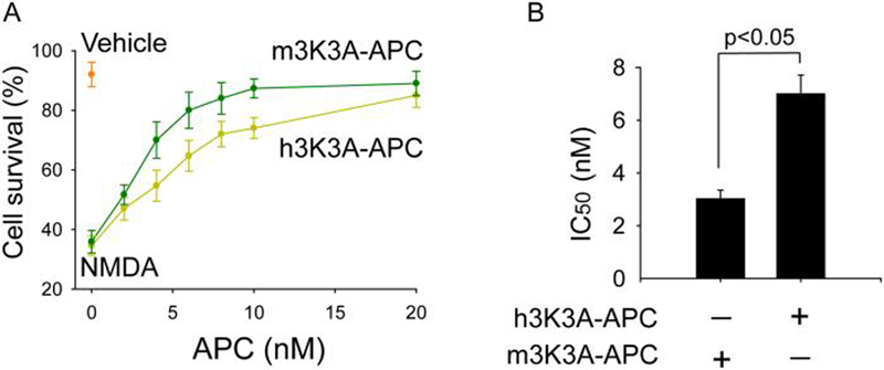
Figure 6. Protection of mouse cortical neurons from NMDA-induced injury by mouse and human 3K3A-APC.
(A) Dose-dependent neuroprotection by mouse and CHO-derived human 3K3A-APC at 24 h of NMDA. Cell survival was quantified with WST-8 assay. (B) IC50 values for mouse and human 3K3A-APC from dose-response curves shown in Fig. 6A. Values are mean ± SEM, n=3-5 independent experiments in triplicate. NMDA label at zero concentration APC on the abscissa shows cell survival of neurons treated with NMDA only without 3K3A-APC. Vehicle denotes the basal cell survival rate of neurons in the culture medium in the absence of NMDA.

