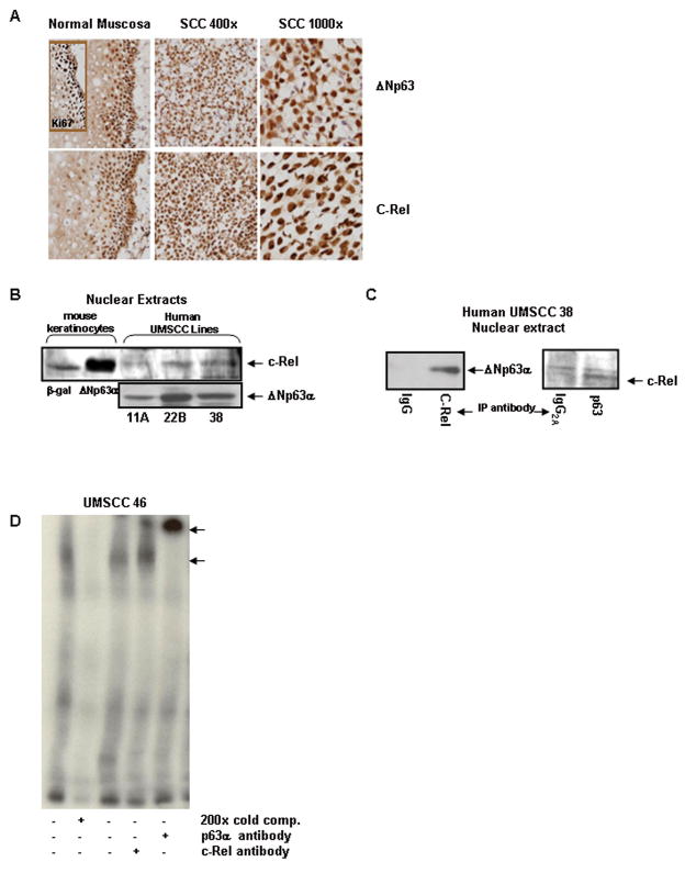
Figure 6. Endogenous ΔNp63α and c-Rel expression are correlated and expanded in primary human cancers, and associate in the nuclei of human squamous cell carcinoma cells.
A) Nuclear expression patterns of ΔNp63α and c-Rel are expanded and associated in primary human squamous cell carcinomas. Immunostaining of normal mucosa and squamous cell carcinoma tissue sections with p63 and c-Rel. The proliferative compartment of normal mucosa is identified by Ki67 immunoreactivity. B) Endogenous Δ Np63α and c-Rel are present in nuclei of human HNSCC lines. Western blots of nuclear extracts prepared from SCC squamous cell carcinoma lines. Mouse keratinocytes overexpressing ΔNp63α or β-gal are included as controls. C) Endogenous nuclear ΔNp63α and c-Rel physically interact in squamous cell carcinoma cells. Co-immunoprecipitation analysis of UMSCC-38 nuclear extracts. Nuclear extracts were immunoprecipitated with antibody to c-Rel (left panel) or p63 (right panel) and probed for c-Rel or p63, as noted. D) Endogenous nuclear ΔNp63α and c-Rel derived from squamous carcinoma cell lines are associated on the p21WAF1 promoter in vitro. EMSA analyses of nuclear extracts derived from the HNSCC cell line UMSCC 46. A 32P labeled probe using the p63 binding site #1 from the p21WAF1 promoter was used in these reactions. A protein:DNA complex is seen and can be partially supershifted with a c-Rel antibody. Use of the smaller 32P tag in the HNSCC experiments allowed for a supershift band to be seen with the p63 antibody as well.

