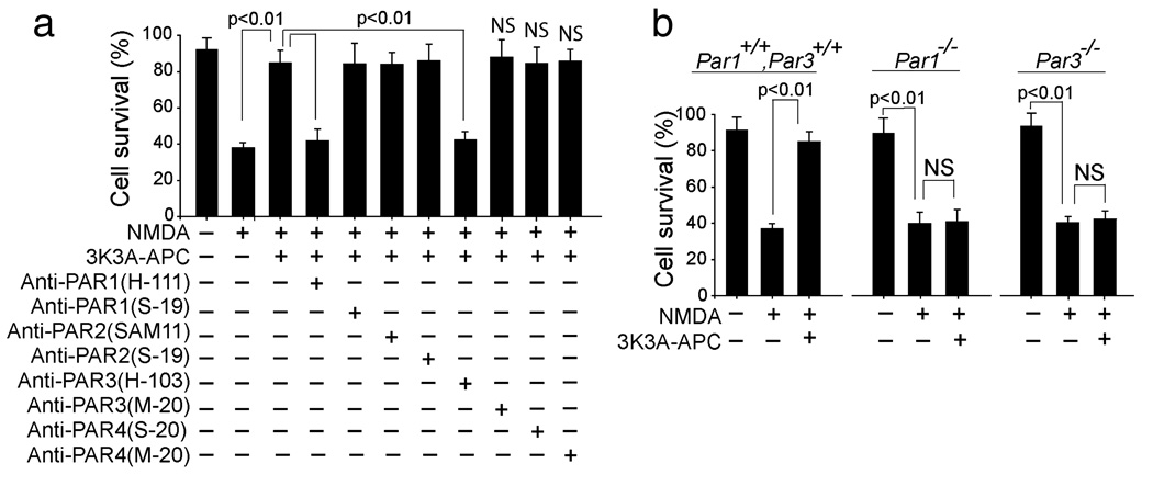
FIG. 3.
The neuroprotection of 3K3A-APC in NMDA-treated neurons is mediated by PAR1 and PAR3. (a) Cortical neurons treated with NMDA and incubated with recombinant murine 3K3A-APC (5 nm and various cleavage-site-blocking antibodies (20 µg/mL) that specifically block the actions of PAR1 (H-111), PAR2 (SAM-11), PAR3 (H-103) and PAR4 (S-20). N-terminal-blocking PAR1 (S-19) and PAR2 (S-19), and C-terminal-blocking PAR3 (M-20) and PAR4 (M-20) antibodies (20 µg/mL) were used as negative controls. Cell survival was quantified with WST-8 assay as in Fig. 1b. (b) Cortical neurons from PAR1−/− and PAR3−/− mice were treated with NMDA and incubated with and without 3K3A-APC (5 nm). Cell survival was quantified with a WST-8 assay. All values are mean + SEM. NS, non-significant.

