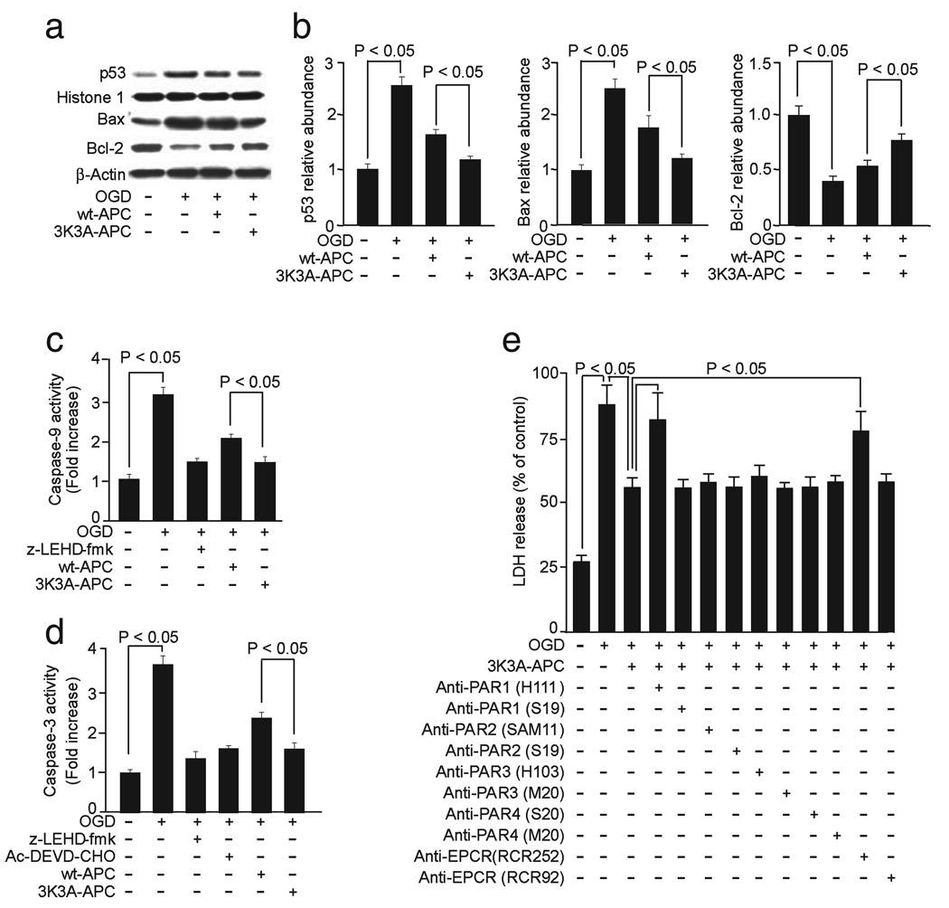
FIG. 6.
Effects of human 3K3A-APC on activation of caspases-9 and -3, p53 levels and Bax and Bcl-2 expression and requirements for EPCR and PAR1 in hypoxic human BECs. (a) Western blots for p53 in nuclear extracts, and Bax and Bcl-2 in whole-cell extracts from BECs treated with OGD for 3:00 h and incubated with and without human 3K3A-APC (10 nm). Histone H1 was used as a control for loading of nuclear p53 protein and β-actin as a control for loading of the whole-cell lysate proteins. (b) Intensity of p53, Bax and Bcl-2 signals measured by scanning densitometry and effects of human 3K3A-APC in experiments as in (a). Signal for nuclear p53 was normalized by histone H1 abundance, and signal for Bax and Bcl-2 with β-actin. Caspase-9 (c) and caspase-3 (d) activities in OGD BECs with and without caspase-9 inhibitor (10 µm; z-LEDH-fmk) and/or caspase-3 inhibitor (50 µm; Ac-DEVD-CHO) and human 3K3A-APC (10 nm). (e) BECs were treated with OGD for 8:00 h and incubated with human 3K3A-APC (10 nm) and various cleavage-site-blocking PAR1 (H-111), PAR2 (SAM-11), PAR3 (H-103) and PAR4 (S-20) antibodies (20 µg/mL) and antibodies against EPCR that either block (RCR252) or do not block (RCR92) APC binding. N-terminal-blocking PAR1 (S-19) and PAR2 (S-19) antibodies and C-terminal-blocking PAR3 (M-20) and PAR4 (M-20) antibodies (20 µg/mL) were used as negative controls. BEC injury was quantified with LDH assay as in Fig. 5b. All values are mean + SEM.

