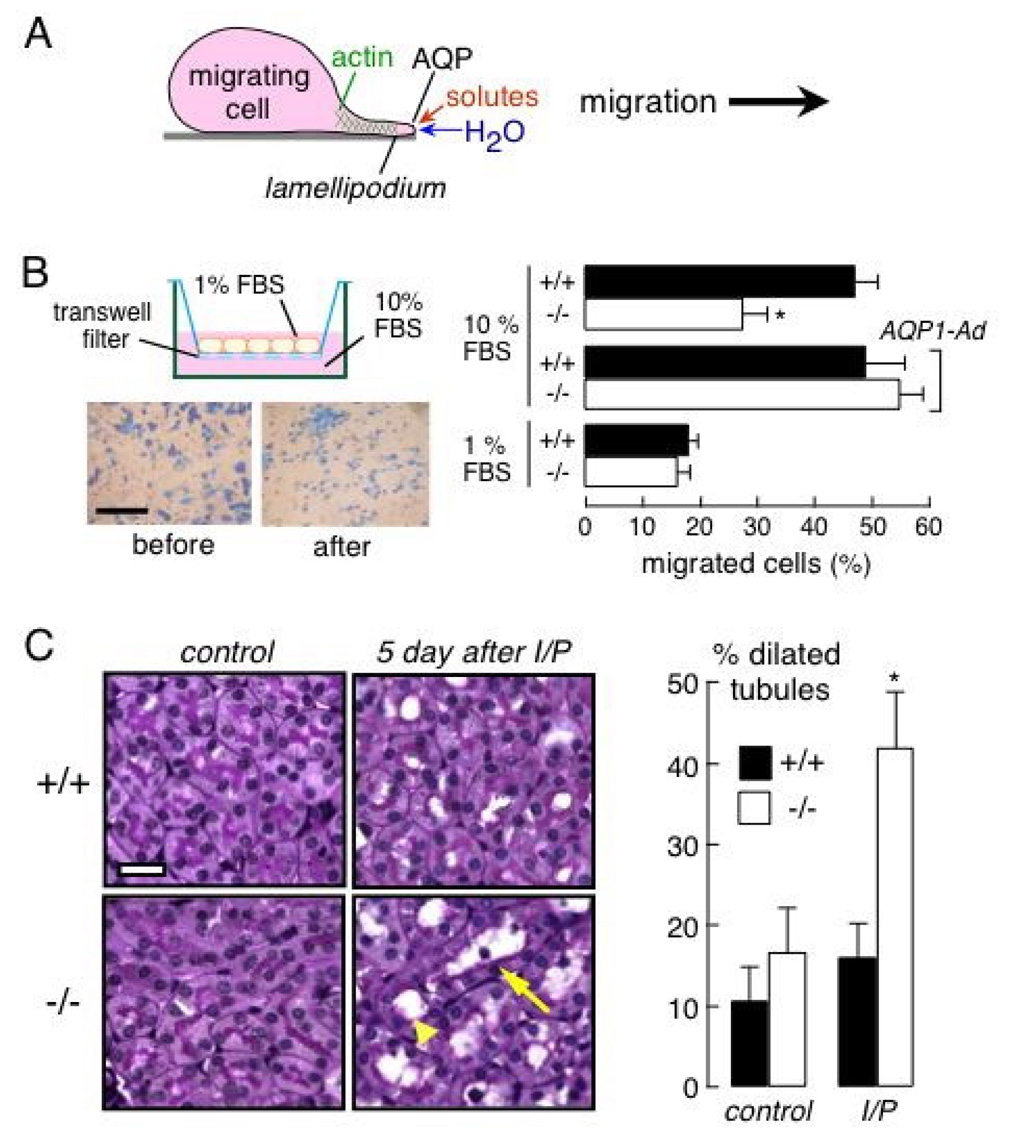
Figure 4. AQP1-facilitated migration of proximal tubule epithelial cells.

A. Proposed mechanism for AQP-facilitated cell migration showing water transport into lamellipodia at the leading edge of migrating cells. See text for further explanations. B. (left) In vitro Boyden chamber assay of proximal tubule cell migration. (top) Schematic of modified Boyden chamber containing a transwell porous membrane filter. Cells migrate from top to bottom in response to a chemotactic stimulus. (bottom) Photographs of adherent proximal tubule cells at the completion of an assay, shown before scraping (left), and after scraping the upper surface (right) to visualize migrated cells. Bar, 100 µm. (right) Percentage of migrated cells at 6 h after cell plating (S.E., * p < 0.01). The bottom chamber contained 10% or 1% FBS. ‘AQP1-Ad’ indicates adenoviral-mediated AQP1 gene delivery. C. Ischemia-reperfusion model of acute renal tubular injury. (left) Periodic acid-Schiff and hematoxylin staining of kidney sections from control wildtype and AQP1 null mice (left), and at 5 days after ischemia/reperfusion (I/P) (right). Bar, 20 µm. Arrowhead, dilated tubule; arrow, degenerated tubule. (right) Percentage of proximal tubules with inner diameter greater than 5 µm (S.E., * p < 0.05). Adapted from ref. 41.
